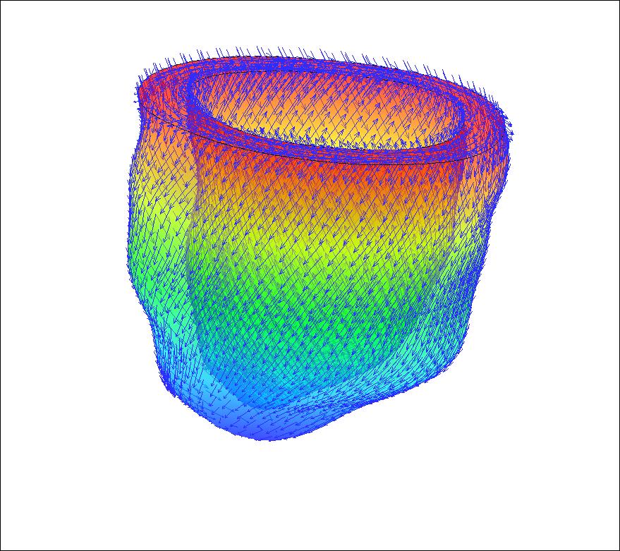
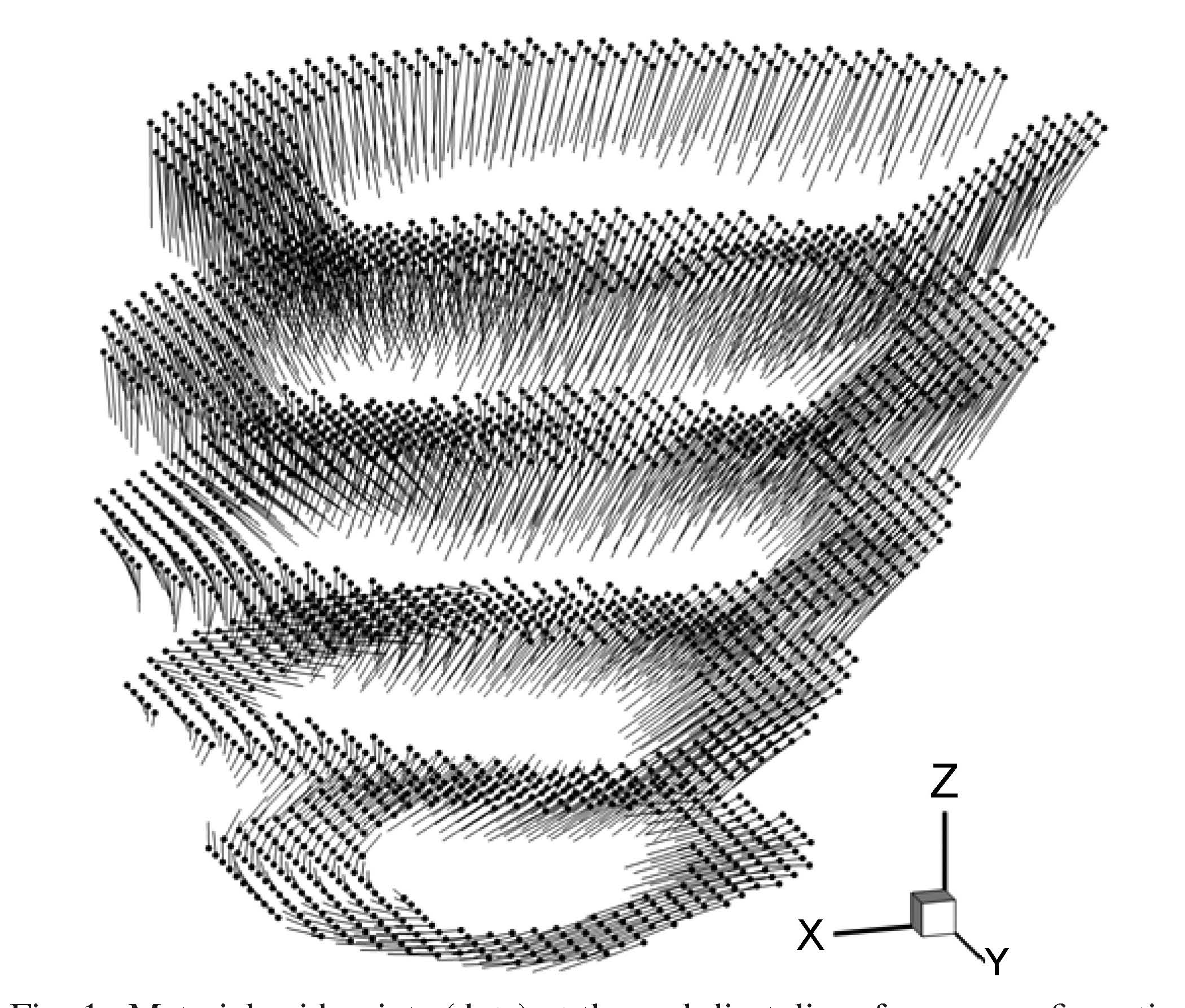
Human left ventricle model rebuilt from MRI images with fibre directions www.GlasgowHeart.org.
Heart attack is a leading cause of premature ill health and death and places a significant burden on the NHS. Consequently, improved approaches are urgently needed to identify the extent of heart muscle injury and potentially predict future outcomes, including treatment response.
Our work embraces this problem by bringing together imaging, mathematics and medicine. We use novel methods with magnetic resonance imaging (MRI) and mathematics to construct 3D multi-scale modelling of heart, with
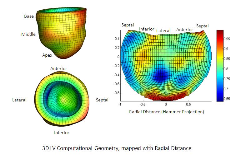
intention to explore acute myocardial infarction (MI) in experimental animals and humans. The cardiac MRI methods provide information on structure, function and pathology in a single scan. The mathematical methods take the MRI data to recreate geometric models of the heart, include fibre structure, and making use of structure-based constitutive models and bi-domain based active contraction. The parameters of the constitutive models will be identified using an inverse approach from the dynamic strain field measured by the 3D displacement encoding with stimulated-echos (DENSE) MRI from human subjects at the Glasgow BHF centre. Since MRI is safe and can be repeated, heart models can be developed serially and so evolve in response to changes in the heart post-MI.
For the mechanical modelling of the heart, we use the the finite-element immersed boundary method - IBAMR, modelling the 3D finite strain with fluid-structure interaction. IBAMR combines the strength of the
conventional IB methods and the finite element methods, and enables both the fluid-structure interaction, and the complicated nonlinear finite strain solid mechanics to be modelled accurately and effectively.
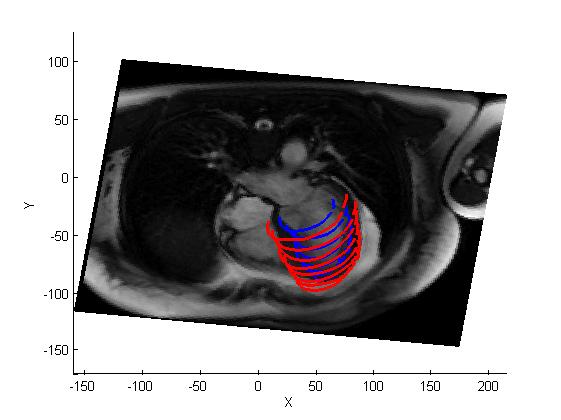
3D strain field measured by DENSE (left), and the automatic geometry extraction of the left ventricle from MRI (right).
The project is funded by the EPSRC, the Medical Research Scotland, and the British Heart Foundation.
Current Research Fellows:
Current PhD students:
Move up to Prof. X. Y. Luo